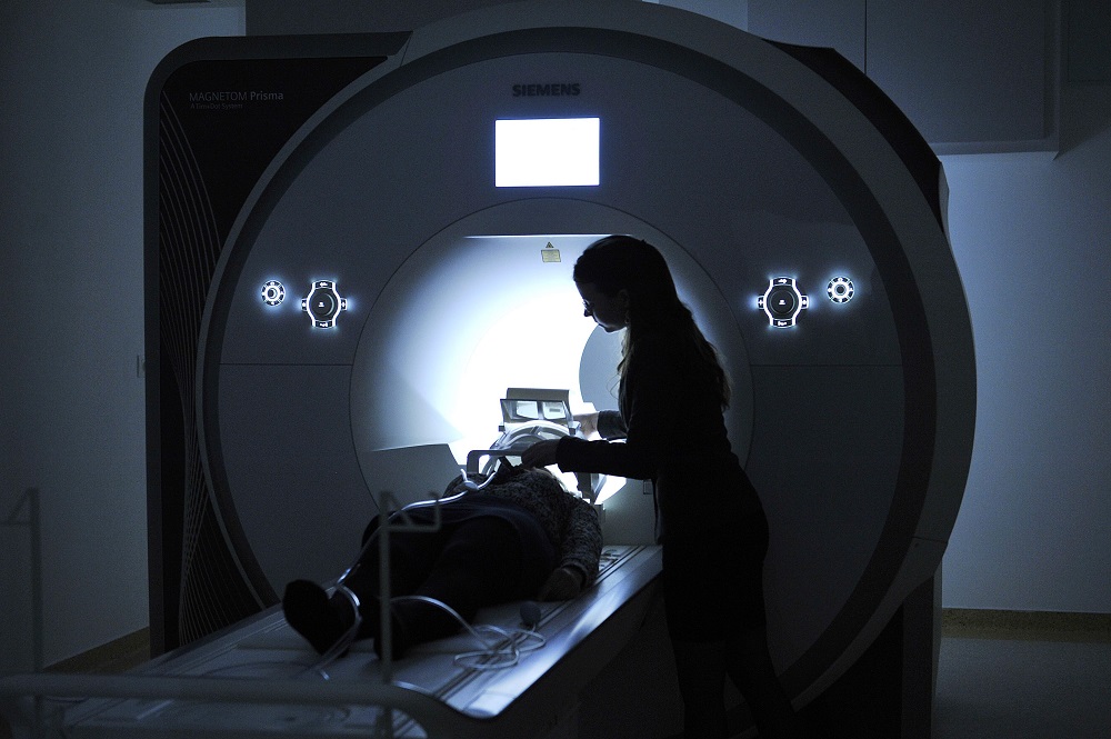
Artificial intelligence-based solutions play an increasingly important role in the analysis of medical images. These systems may support clinical decision making and biomedical research as well. Quality control of the obtained images is of paramount importance for both scientific research and clinical applications. Automating the quality control of MRI scans can save significant time and money while increasing the reliability of the results.
State-of-the-art image recognition technologies use artificial neural networks based on deep learning to analyze images. However, a difficulty in the field of MR image quality control is that expert-rated images are usually not available in sufficiently large quantities to allow the effective training and application of such complex networks. In such data-poor environments, traditional machine learning methods based on predefined image features are available as an alternative option. However, the disadvantage of these methods is the significant processing time required to extract the image features when compared to neural networks that are capable of making a decision about an image in a fraction of a second.
In the present study, researchers at the Brain Imaging Centre of RCNS performed a systematic comparative analysis to investigate the extent to which similar performance can be achieved with deep learning-based methods when compared to machine learning models based on standard image features.
The authors of the study used publicly available brain imaging data as well as data collected in the Brain Imaging Centre MR laboratory. The quality of the images was assessed by a team of expert radiologists from the point of view of clinical diagnostic utility. A low-complexity artificial neural network was developed that learned to classify the images according to whether they are suitable for identifying brain abnormalities or not, based on the expert ratings. The network performed exceptionally well in testing, classifying previously unseen images with an accuracy over 94%. Similar classification performance was achieved with classical machine learning models using image-derived features on the same task.
Of particular importance for the field as a whole is that the research has demonstrated the suitability of deep learning-based methods for the rapid evaluation of brain MR image quality from a clinical point of view. The developed deep learning model offers promising new possibilities for automating quality control in other areas of medical imaging as well, thus facilitating efficient clinical decision making.

The results of the RCNS Brain Imaging Centre, achieved with the support of GINOP and ERANET (GINOP-2.2.1-18-2018-00001, 2019-2.1.7-ERA-NET-2020-00008) grants, have been published in the most prestigious journal in the field, Medical Image Analysis.
Pál Vakli*, Béla Weiss*, János Szalma, Péter Barsi, István Gyuricza, Péter Kemenczky, Eszter Somogyi, Ádám Nárai, Viktor Gál, Petra Hermann, Zoltán Vidnyánszky, 2023. Automatic brain MRI motion artifact detection based on end-to-end deep learning is similarly effective as traditional machine learning trained on image quality metrics. Medical Image Analysis, https://doi.org/10.1016/j.media.2023.102850

